Novtember 11, 2022

Stuttering, basal ganglia, and cerebellum

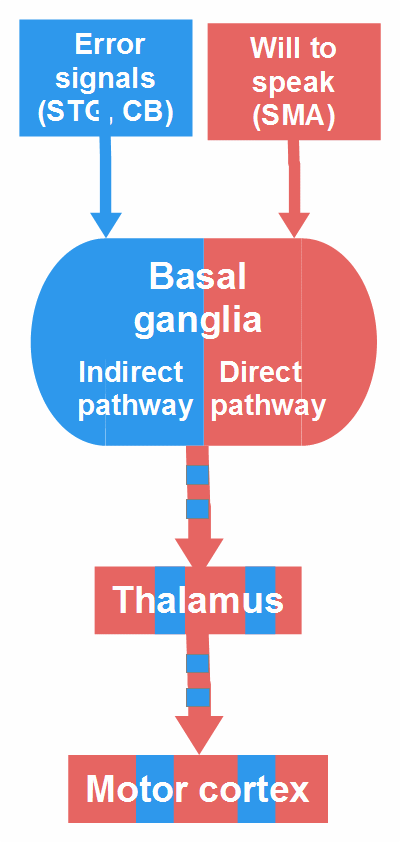
In July 2019, I posted a figure (Fig. 21) in which two components of a stuttering event are symbolized: an ‘impelling’ component (depicted in red) representing the person’s will to speak, and an ‘inhibiting’ component (depicted in blue) that interrupts the flow of speech against the person’s will. The basal ganglia are part of the first component, the cerebellum is part of the second one in that figure. This view was influenced by the ‘dual premotor model’ proposed by Goldberg (1985) and applied to stuttering by Alm (2006, 2007). The model reflects the traditional assumption that basal ganglia and cerebellum are anatomically separate subcortical systems that perform unique functional operations and interact only at the level of the cerebral cortex.

This view is obsolete; studies with animals and humans found that cerebellum and basal ganglia interact at the subcortical level and form an integral network (Bostan & Strick, 2018; Wang et al., 2020). Thus, I had to once more consider the role of basal ganglia and cerebellum in stuttering, and in this post I will present what came out. I still maintain the view that stuttering is caused by invalid error signals in the monitoring system – ‘invalid’ in the sense that no speech error happened – and that the cerebellum is critically involved in the generation of these error signals (see Section 2.1 in the main text). However, the role of the basal ganglia (BG) is more complicated than I assumed earlier: They are not a combatant in the struggle between the two components of stuttering, they are the battlefield.

For a long time, I believed I would never understand the intricate BG mechanism (and I think it is not yet completely understood by the experts), but I got that, in terms of motor control, there are two main pathways through the BG: a so-called direct pathway that facilitates the execution of a planned voluntary motor action, and a so-called indirect pathway that inhibits a motor action (details below). These two pathways operate like two reins, one for Go and one for NoGo. The prevalence of the one or the other pathway decides on what we do or not do. Both pathways are modulated by inputs from many cortical and subcortical areas, such that knowledge, experience, and inherent instincts can influence the decision in the BG. For instance, the direct pathway gets support by the nucleus accumbens whose activity is associated with the expectation of reward, whereas the indirect pathway is supported by the amygdala whose activity is associated with fear.
Is the hyperdirect pathway involved in stuttering?

In addition to the direct and the indirect pathway, there is a ‘hyperdirect pathway’, so named because it connects the frontal cortex through the subthalamic nucleus (STN) with the globus pallidus pars interna (GPi), the main output structure of the motor part of the BG, without involving the striatum, the usual BG input structure. The hyperdirect pathway was found in studies using Go/NoGo tasks and serves for a global (non-selective) response inhibition. It allows the volitional stopping of an already initiated action, e.g., if the initiation was premature or erroneous (Wiecki & Frank, 2013).

In my penultimate blog post about Martin Sommer’s report of a case in which lifelong stuttering disappeard after a cerebellar damage (see here), I mentioned the hypothesis by Neef et al. (2016, 2017) that the hyperdirect pathway may be involved in stuttering. Neef et al. (2017) write that “stuttering might be caused by an overly active global response suppression mechanism […] the stopping of an ongoing speech motor program and/or the selection of a succeeding speech motor program might fail.”

In my view, this hypothesis is not convincing for three reasons: First, global response inhibition via the hyperdirect pathway is a volitional act, but stopping of an ongoing speech motor program and selection of a succeeding one might be automatic and controlled by the indirect and direct BG pathway, respectively. Second, an overly active global response suppression mechanism would affect not only speaking – or one has to explain why it is overly active during speech only. Third, the hypothesis does not include the cerebellum, although it might play a crucial role in stuttering.

The over-activation of right BA44 in stuttering (a reason for the hypothesis in question) may be related to the attempt to overcome the inhibition of speech, that is, it may not be related to the hyperdirect, but to the direct pathway. I agree with Neef and colleagues when they assume that global response inhibition is involved in stuttering; however, I think it is not evoked by input from the frontal cortex, i.e., not via a hyperdirect pathway, but by input from the cerebellum.

The two components of stuttering and the role of the BG

Two components of stuttering Figure 22: Two components of stuttering (update). STG = superior temporal gyrus, CB = cerebellum, SMA = supplementary motor area.

Figure 22 is an update of the bottom part of Figure 21. I retain the symbolic colors: red for the impelling and blue for the inhibiting component. As mentioned above, the BG are part of both components. In fluent speech, the direct pathway facilitates the execution of selected speech motor programs. A stutter occurs when, by cerebellar input, the indirect pathway becomes transiently dominant and inhibits the execution of the selected or initiated speech motor program, whereas the speaker tries to continue. This attempt (= input from the frontal cortex) activates the direct pathway, and this activation contributes to overt stuttering symptoms: They would not occur, if the speaker, at the moment of inhibition, could give up the will to speak.

As mentioned, Figure 22 updates only the bottom part of Figure 21; regarding the upper part, I still think that error signals in the monitoring system (possibly a loop connecting superior temporal cortex, cerebellum, and thalamus) are caused by poor processing of auditory feedback as a result of a misallocation of attention during speech. An error signal at speech onset may occur when a speech motor program is initiated before the entire speech network, including attention and auditory system, is sufficiently prepared – or specifically, when speaking starts without any expectation of auditory input. The start may then be regarded as premature by the monitoring system and inhibited by the BG.

The direct pathway and dopamine

Stuttering theory, basal ganglia and stuttering Figure 23: Direct pathway. Arrows symbolize only the direction of the projections. DA = dopamine, GPi = globus pallidus pars interna, SMA = supplementary motor area, SNc = substantia nigra compacta, SNr = substantia nigra reticulata.

Figure 23 shows the direct pathway through the BG. The frontal cortex, particularly the SMA, excites ‘Go neurons’, specific medium spiny neurons in the dorsal striatum, that inhibit GPi and SNr, the output structures of the BG. Since they themselves have inhibitory projections to the thalamus, the result is a disinhibition, i.e., an excitation of the thalamus, which excites the motor cortex and facilitates the execution of the selected motor program. (Bostan & Strick, 2018; Wiecki & Frank, 2013).

The direct pathway is reinforced by dopaminergic projections from the substantia nigra compacta (SNc) and the nucleus accumbens (part of the ventral striatum, not depicted in Fig. 23) to the dorsal striatum. Via D1 receptors, dopamine further excites active Go cells in the striatum, via D2 receptors, dopamine inhibits NoGo cells (Wiecki & Frank, 2013). In this way, dopamine supports the drive of habitual, automatic, promising behavior, the impact of motivation and of expected success or reward.

In stuttering, the role of dopamine may be ambiguous: It supports fluent speech, but when speech flow is interrupted for no clear reason, high dopamine reception in the striatum may drive effort and struggle behavior, thereby increasing the severity esp. of tonic stuttering. Studies showed that the nucleus accumbens in the right hemisphere is larger in adults who stutter than in controls (Neef et al., 2018; Montag et al., 2019). Further, Watkins et al. (2008) and Metzger et al. (2018) found a positive correlation between substantia nigra activity and stuttering severity, in the latter case during a non-speech task.

The BG are not only involved in motor control but also in attention regulation, not least because voluntary actions are always associated with selective (top-down) attention. A generally high dopamine level in the striatum might support focused attention and hamper distraction, but also attention shifting, and attention distribution. This corresponds to behavioral findings showing that stuttering children and adults performed poorer on average than controls in Go/NoGo and dual tasks (see Section 3.3.1 in the main text). This is no BG dysfunction, but a personality trait. A strong propensity to goal-directed activity and focused attention is advantageous in not a few jobs, but it may increase the risk of stuttering.

The indirect pathway and the role of the cerebellum

Different from the direct pathway that facilitates and ‘releases’ voluntary movements, the basic function of the indirect pathway is the selective inhibition of concurrent unwanted, countering movements. For example, we cannot concurrently stretch and flex a limb. A further function of the indirect pathway is the global inhibition of premature, erroneous, or potentially dangerous actions. Global inhibition can be elicited volitionally, that is, by input from the frontal cortex to the STN (hyperdirect pathway, see above), but I think it can be elicited also involuntarily and even against the will, namely by input from subcortical structures such as the cerebellum, the amygdala, or the periaqueductal gray (the activity of the latter is associated with fury). Studies in humans and animals show that these structures are interconnected with the STN (Accolla et al., 2016; Hachem-Delaunay et al., 2015; Smith, Hazrati, & Parent, 1990).

Stuttering theory, basal ganglia, cerebellum Figure 24: Indirect pathway and the cerebellum-BG loop. Arrows symbolize only the direction of the projections. DN = dentate nucleus, GPe = globus pallidus pars externa, GPi = globus pallidus pars interna, ITN = intralaminar thalamic nuclei, PN = pontine nuclei, SMA = supplementary motor area, SNr = substantia nigra reticulata, STG = superior temporal gyrus, STN = subthalamic nucleus. The figure does not respect spatial relations; for instance, the ITN, of course, are part of the thalamus.

Normally, in case of selective inhibition, the cortex sends excitatory projections to NoGo cells in the dorsal striatum (this is not depicted in Fig. 24, but imagine the red boxes and arrows in the figure were blue). The striatal NoGo cells send inhibitory projections to the GPe, and the GPe has inhibitory projections to the STN; thus the inhibition of the GPe disinhibits, i.e., activates the STN. Now, the STN, by excitatory projections, activates GPi and SNr, the BG output structures, which inhibit thalamus and motor cortex.

Figure 24 shows what may happen in stuttering: The cortex sends excitatory projections to Go cells in the striatum (representing the will to speak) and, just as in the direct pathway, these cells try to inhibit the GPi (this is symbolized by the red boxes and arrows). But now, the loop between BG and cerebellum comes into play. This loop interconnects the monitoring system (STG, cerebellum, thalamus) with the ‘executive system’ (SMA, BG, thalamus, motor cortex). When a motor sequence such as speech is performed, the STN permanently ‘asks’ the cerebellum whether each segment of the sequence (each word, syllable, or phrase) was properly produced. The cerebellum, or a loop between cerebellum and STG, compares auditory predictions with auditory feedback. If a mismatch is detected, the cerebellum sends input to NoGo cells in the striatum. This activates the indirect pathway that takes over: As depicted in Fig. 24, the GPe is inhibited, this activates the STN that excites GPi and SNr, and they inhibit the thalamus and the motor cortex.

This view is consistent with Metzger et al, (2018) who observed an increased functional coupling between GPe and cortex, suggesting “an excessive activity of the indirect pathway and, thus, an increased inhibitory action on the cortex during stuttering, which is in line with previous reports of the fluency-enhancing effect of D2 receptor blockers” (p. 177). The empirical basis for the proposed scenario comes from studies in mammals and humans in which BG and cerebellum were found to form an integral network (for an overview, see Bostan & Strick, 2018). The STN is the source of a dense disynaptic projection to the cerebellar cortex (lobule VI, crus I/II) via the pontine nuclei, and the dentate nucleus in the cerebellum is the source of a dense disynaptic projection to the striatum via the intralaminar thalamic nuclei (Bostan & Strick, 2018; Wang et al., 2020). The cerebellar output targets NoGo neurons in the striatum, that is, it preferentially influences the indirect pathway (Bostan & Strick, 2018).

Not a few studies have shown the cerebellum to be involved in the detection and correction of motor errors, based on sensory feedback (see footnote in the main text). For instance, Zheng et al. (2013) identified a brain network that appeared to encode an error signal in response to distorted auditory feedback during articulation. Seidler, Noll, and Chintalapati (2006) found BG activation in a task involving error-based sensorimotor adaptation. Bostan and Strick (2018) assume that the pathway from the cerebellum to the striatum underlies this activation.

In stuttering, inhibition of the motor cortex is possibly not the most important effect of the STN activation. Inhibition of the motor cortex would only mean that muscles are no longer activated. But the sudden stopping of a current or initiated action requires either the activation of antagonistic muscles (stopping the current movement) or the prolongation of current muscle contraction (making the transition to the next movement impossible). Just this happens in tonic stuttering (for repetitions, see here in Section 2.1). The muscle contractions could possibly be elicited via a pathway from the STN to the brainstem. As mentioned, the projection from the STN to the cerebellum is mediated by the pontine nuclei. According to Marien (in Manto et al., 2012), two distinct neural networks control speech sound production: a ‘linguistic’ network (involving Broca’s area, SMA, anterior insula, BG and cerebellum) and, more interesting in our context, a phylogenetically ancient motor pathway for vocalization, which encompasses, among others, BG and pons and projects to cranial nerve motor nuclei in the lower brainstem that are responsible for the innervation of the vocal tract musculature. This pathway from the BG to the brainstem could interrupt speech flow in stuttering.

The involvement of that subcortical motor pathway for vocalization would account for the fact that, in stuttering (except word repetition), typically the vowel of a syllable cannot be produced (Wingate, 1988): If a syllable starts with a vowel, vocalization is inhibited at syllable onset (silent block); in the other syllables, the transition to the vowel is hampered by repetition or prolongation of the preceding consonant(s). The activity of a BG-brainstem pathway (without cortical involvement) would also account for the subjective experience of stuttering: that a power stronger than the will, for reasons inaccessible to consciousness, suddenly interrupts speaking.

Global response inhibition

As already mentioned, I think that the overactivation of the STN-cerebellum loop following an invalid error signal causes a global inhibition; that is (acc. to Wiecki & Frank, 2013), the STN, by excitatory projections to the SNr, enhances the threshold for any action, independent of action modality. This would explain the reduction of stuttering frequency when the natural speech rhythm is accompanied by significant gestures, by hand-, arm-, or other bodily movements. Global inhibition is then more difficult because the additional movement would have to be inhibited too. But there is no reason to inhibit this movement, as it doesn’t cause error signals, and so it strengthens the direct BG pathway.

A further argument for global response inhibition being the immediate cause of stuttering is the fact that a stutter block becomes weaker after a short time, such that the speaker can continue (until the next block occurs). This corresponds to the course of global response inhibition: When the STN is over-activated, it sends excitatory projections also to the GPe (this is not depicted in Fig. 24). Since the GPe has inhibitory projections to the STN, the excitation of the GPe, after a delay, inhibits the STN, such that the global NoGo signal becomes weaker and disappears. Then, the direct pathway takes over and allows the selected motor program to be executed (Frank, 2006; Wiecki & Frank, 2013).

One may ask: Is it likely that cerebellar output interferes in such a severe way with the process in the BG? An example for cerebellar impact on the BG is dystonia. Similar to stuttering, dystonia has often been considered a BG disorder, but growing evidence suggests that the cerebellum is involved. In animal models, dystonic postures were evoked by abnormal cerebellar output and abolished by silencing or lesioning the cerebellum (Bostan & Strick, 2013; Tewari, Fremont, & Khodakhah, 2017). However, whereas dystonia seems to be caused by cerebellar dysfunction, the cerebellum might do its job in stuttering: it responds to error signals in the monitoring system – these error signals are the problem.

The involvement of the cerebellum in stuttering is strongly suggested by cases in which lifelong stuttering disappeared after impairment of the cerebellar function (Miller, 1985; Bakheit, Frost, & Ackroyd, 2011; a further case was in Germany in 2012, see here). In our context, a case reported by Muroi et al. (1999) is particularly interesting: A 66-years-old man lost his lifelong severe stuttering after a stroke that affected the medial thalamus bilaterally. The patient not only lost his stuttering, his personality regressed to that of an approximately 10 year-old boy. Before the stroke, he was a serious, “hard-grained”, taciturn man, after awakening from the coma, he was easy-going and showed childish behaviors, without other neuropsychological dysfunctions. Possibly, cerebellar output could no longer influence the process in the BG because the pathway to the striatum was interrupted after the lesion in the medial thalamus; therefore, signals from the cerebellum could neither inhibit speaking nor inappropriate, childish behaviors.

At the end, I want to emphasize that the above – except the empirical findings I refer to – is hypothetical and in part speculative. My aim was: to show a possible way how invalid error signals occurring in the monitoring system can cause stuttering. But as far as I see, the proposed hypotheses is coherent in itself and consistent with the relevant data.

to the top

previous page